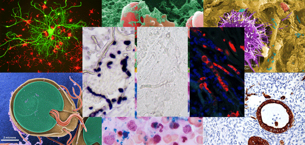
It is known that in-situ hybridization can be used to determine the cryptologic and histological location of the nucleic acid of interest
Fluorescent in Situ Hybridization (FISH)
Probe is a subatomic cytogenetic technique that visualizes genetic materials
using fluorescent probes. These probes are molecules that absorb a specific
wavelength of light and emit light when they bind to a specific DNA/RNA
sequence. They are used to detect structural and numerical chromosomal
abnormalities, monitor therapeutic drugs, and identify rare genetic diseases.
FISH probes that are commonly used include locus-specific, Alphoid/centromeric
repeat, and whole chromosome probes. They have several advantages, including
high sensitivity and accuracy in recognizing targeted sequences, direct
application to both metaphase and interphase nuclei, and accurate visualization
of hybrid signals at the single-cell level.
One of
the key factors driving the market growth is the increasing prevalence of
various genetic and chronic disorders. Furthermore, the rising demand for In
Vitro Diagnostics (IVD) testing and targeted therapies around the world is
propelling the market forward. When compared to traditional cytogenetic (cell
gene) tests, FISH tests can detect minute genetic changes that are normally
missed under the microscope. As a result, these probes are widely used for
cancer and genetic disorder diagnosis, prediction of outcomes, and clinical
management. Furthermore, various technological advancements, such as the
development of FISH probes with higher sensitivity and accuracy, are
contributing to the market's positive outlook. Other factors, such as improving
healthcare infrastructure, particularly in developing countries, and extensive
research and development (R&D) in biotechnology, are expected to drive the
market even further.
In-situ Hybridization (ISH) is a
well-established set of methods for detecting and visualizing specific nucleic
acid sequences in whole organisms, cytological preparations, and tissue
sections. Over the several decades of its use, the technique has seen
refinements and a significant increase in applications, and it is still
routinely used in laboratories where visualization of gene expression within
the tissue of concern is required.
In-situ hybridization is known to allow for
the cryptologic and histologic localization of the nucleic acid of interest.
While the techniques have matured significantly in the current scenario, the
application of most In-situ Hybridization techniques
has been severely limited due to their inability to detect samples with low
copies of DNA and RNA. Advances in recent years have resulted in the
development of a number of strategies to help improve the sensitivity of ISH
techniques, such as signal detection after hybridization or amplification of
the target nucleic acid sequence before ISH begins.


Comments
Post a Comment